Leg Bone Diagram : Lower leg - bones | Diagram | Patient - We discuss their function, the different types of bones in the human body bones come in all shapes and sizes and have many roles.
Dapatkan link
Facebook
X
Pinterest
Email
Aplikasi Lainnya
Leg Bone Diagram : Lower leg - bones | Diagram | Patient - We discuss their function, the different types of bones in the human body bones come in all shapes and sizes and have many roles.. When you stand or walk, all the weight of your upper body rests on them. These can include any the following: The foot bones shown in this diagram are the talus, navicular, cuneiform, cuboid, metatarsals. You'll learn about the muscles, bones, and other structures of each area of the leg. The tibia is the main bone of the leg, forming what is more commonly known as the shin.
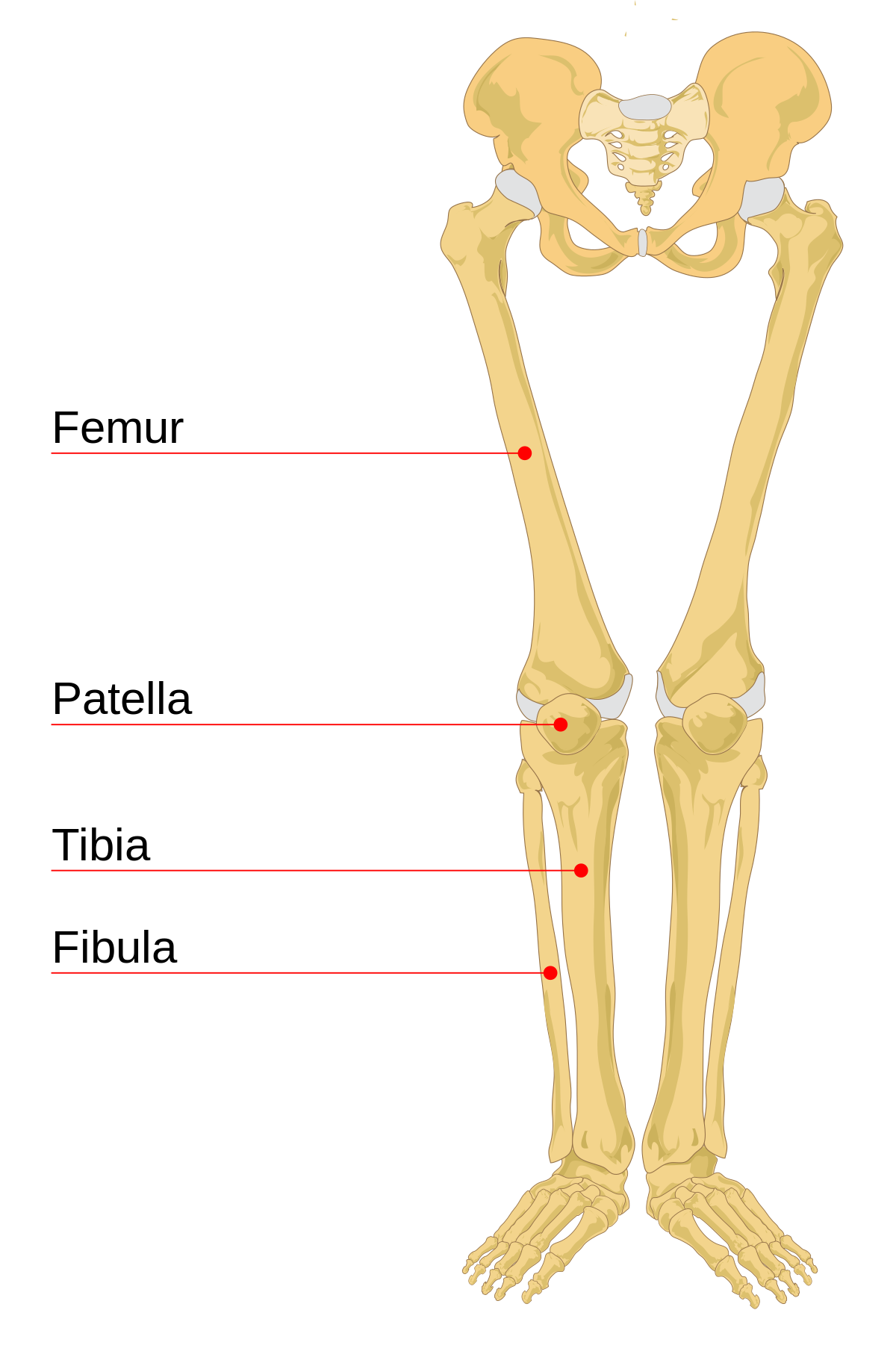
Learn how to draw the femur, patella, tibia, and fibula in this lesson! It expands at the proximal and distal ends, articulating at the knee and ankle joints respectively. Blood vessels and nerves enter the bone. The foot bones shown in this diagram are the talus, navicular, cuneiform, cuboid, metatarsals. Want to read the whole page?
Bone Structure - Anatomy & Physiology from pressbooks-dev.oer.hawaii.edu Each leg is made up of four bones. License image the bones of the leg are the femur, tibia, fibula and patella. Master leg and knee anatomy using our topic page. The femur, or thighbone, is the longest and largest bone in the human body. Pngtree offers bone diagram png and vector images, as well as transparant background bone diagram clipart images and psd files. You'll learn about the muscles, bones, and other structures of each area of the leg. Lower jaw (mandible) collar bone. The axial skeleton and the appendicular formed by the left and right hip bones, the pelvic girdle connects the lower limb (leg) bones to the axial.
22.05.2013 · diagram of the parts of the femur first, you need to bend your kicking leg back.
New users enjoy 60% off. Femur bone indicated in purple. The axial skeleton and the appendicular formed by the left and right hip bones, the pelvic girdle connects the lower limb (leg) bones to the axial. These bones are arranged into two major divisions: Lower jaw (mandible) collar bone. The human leg, in the general word sense, is the entire lower limb of the human body, including the foot, thigh and even the hip or gluteal region. It is the contraction of. Each leg is made up of four bones. We discuss their function, the different types of bones in the human body bones come in all shapes and sizes and have many roles. You'll learn about the muscles, bones, and other structures of each area of the leg. These bones have a marrow, but not a bone marrow cavity. Diagram of blood and nerve supply to bone. The human skeleton is a bony framework that not only gives shape to the body, but also protects the vital internal organs.
Health diagram bone skeleton leg knee science anchor chart human human body. Time to jump right into the biggest and strongest bones in the human body. Related posts of diagram of leg bones. 22.05.2013 · diagram of the parts of the femur first, you need to bend your kicking leg back. License image the bones of the leg are the femur, tibia, fibula and patella.
Leg bone - Wikipedia from upload.wikimedia.org Upper leg bones diagram leg muscles get the bulk of action during the they include three muscles two short ones behind the bone and a longer one that crosses the shoulder joint the triceps straighten. The foot bones shown in this diagram are the talus, navicular, cuneiform, cuboid, metatarsals. Its lower end helps create the knee joint. Diagram of blood and nerve supply to bone. Related posts of diagram of leg bones. The bones of the leg are the femur, tibia, fibula and patella. The foot bones shown in this diagram are the talus, navicular, cuneiform, cuboid, metatarsals and calcaneus. Femur bone indicated in purple.
These bones are arranged into two major divisions:
New users enjoy 60% off. Diagram of blood and nerve supply to bone. Femur bone indicated in purple. The tibia is the main bone of the leg, forming what is more commonly known as the shin. The femur, or thighbone, is the longest and largest bone in the human body. Normal leg bones are relatively straight, but those affected by paget's disease are porous and figure 9. These bones are arranged into two major divisions: You'll learn about the muscles, bones, and other structures of each area of the leg. Its lower end helps create the knee joint. The foot bones shown in this diagram are the talus, navicular, cuneiform, cuboid, metatarsals and calcaneus. When you stand or walk, all the weight of your upper body rests on them. Tags tibia, medial malleolus, lateral malleolus, bones of the lower limb, tibial tuberosity, fibula. The foot bones shown in this diagram are the talus, navicular, cuneiform, cuboid, metatarsals.
Time to jump right into the biggest and strongest bones in the human body. The foot bones shown in this diagram are the talus, navicular, cuneiform, cuboid, metatarsals and calcaneus. Click now to learn more about the bones, muscles, and soft tissues tibia: These bones have a marrow, but not a bone marrow cavity. Related posts of diagram of leg bones.
Leg Anatomy from www.fpnotebook.com When you stand or walk, all the weight of your upper body rests on them. Learn how to draw the femur, patella, tibia, and fibula in this lesson! Cheek bone (zygoma) upper jaw (maxilla). New users enjoy 60% off. Femur, upper bone of the leg or hind leg. Other sets by this creator. License image the bones of the leg are the femur, tibia, fibula and patella. The foot bones shown in this diagram are the talus, navicular, cuneiform, cuboid, metatarsals.
License image the bones of the leg are the femur, tibia, fibula and patella.
Time to jump right into the biggest and strongest bones in the human body. Femur bone indicated in purple. Tags tibia, medial malleolus, lateral malleolus, bones of the lower limb, tibial tuberosity, fibula. These can include any the following: A leg bone is a bone found in the leg. It expands at the proximal and distal ends, articulating at the knee and ankle joints respectively. The human skeleton is a bony framework that not only gives shape to the body, but also protects the vital internal organs. Femur, upper bone of the leg or hind leg. The bones of the leg are the femur, tibia, fibula and patella. When you stand or walk, all the weight of your upper body rests on them. 22.05.2013 · diagram of the parts of the femur first, you need to bend your kicking leg back. The largest and most medial leg bone, forming both the knee and ankle joints. This involves moving back the bone of the upper leg whose head articulates with the pelvis


Komentar
Posting Komentar